09.07.2025
O IGFAE únese a REDONGRA, unha rede para impulsar a ciencia de ondas gravitacionais en España
Novas
Novas científicas

11.03.2021
O Instituto Galego de Física de Altas Enerxías (IGFAE) lidera unha investigación en colaboración coa Universidade de Salamanca na que desenvolven unha nova técnica, baseada en pulsos láser ultracortos, para producir imaxes tomográficas de alta resolución. O traballo publicouse na revista Physica Medica e realizouse no Laboratorio Láser de Aceleración e Aplicacións (L2A2) da Universidade de Santiago
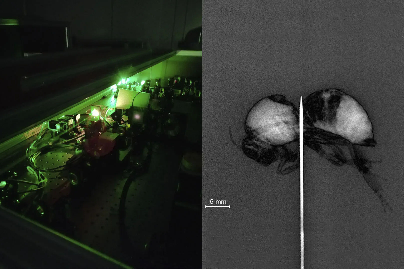
Un equipo liderado polo Instituto Galego de Física de Altas Enerxías (IGFAE), centro mixto da Universidade de Santiago de Compostela (USC) e a Xunta de Galicia, en colaboración coa Universidade de Salamanca, conseguiu xerar raios X mediante unha nova tecnoloxía de aceleración de electróns usando pulsos láser ultracortos. Aínda que antes xa puideran xerar raios X con pulsos láser, esta é a primeira vez que foron producidos de forma estable durante longos períodos de tempo, obtendo as primeiras imaxes tomográficas con esta nova fonte de raios X. Os resultados do traballo foron publicados recentemente no último número da revista Physica Medica.
Esta investigación realízase no Laboratorio Láser de Aceleración e Aplicacións (L2A2) da USC. O L2A2 está equipado cun dos poucos sistemas láser que existen no mundo capaz de producir pulsos láser cunha potencia de 50 teravatios (1012 vatios, un billón de vatios). Ao focalizar estes pulsos sobre un material xéranse campos electromagnéticos moi intensos, capaces de acelerar en poucas micras (0,001 milímetro) partículas a gran enerxía. Esta tecnoloxía permitirá construír aceleradores de partículas compactos con aplicacións médicas no futuro.
“A vantaxe desta nova tecnoloxía é que, ao ter un foco xerador de raios X de dimensión micrométrica, conséguense imaxes de mellor calidade cunha menor cantidade de raios X”, asegura José Benlliure, investigador do IGFAE e coautor deste traballo. “Ademais, pódese aplicar unha nova técnica de reconstrución da imaxe, radiografía por contraste de fase, que permite visualizar materiais de diferente densidade, en particular tecidos biolóxicos”. Os investigadores do IGFAE puideron aplicar esta nova técnica tanto a radiografía convencional como a imaxes tomográficas en tres dimensións.
Recentemente, o IGFAE iniciou un proxecto en colaboración coa Universidade Complutense de Madrid, a Universidade de Salamanca e a empresa Sedecal S.L. para estudar a posibilidade de utilizar esta nova tecnoloxía para desenvolver mamógrafos de alta precisión.
Referencias:
DOI: 10.1016/j.ejmp.2020.12.023
Imaxe: Dereita: vista do pre- amplificador multipaso do láser de L2A2. Dereita: imaxe por contraste de fase dunha abella obtida coa nova fonte microfoco de raios X. Crédito: IGFAE.
Novas